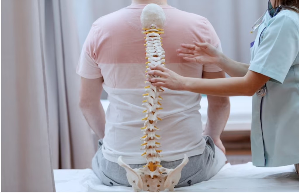
The perceived value of the radiologist to the healthcare team hinges on the accuracy of image interpretation and the quality, clarity, and consistency of the associated report on oncology.
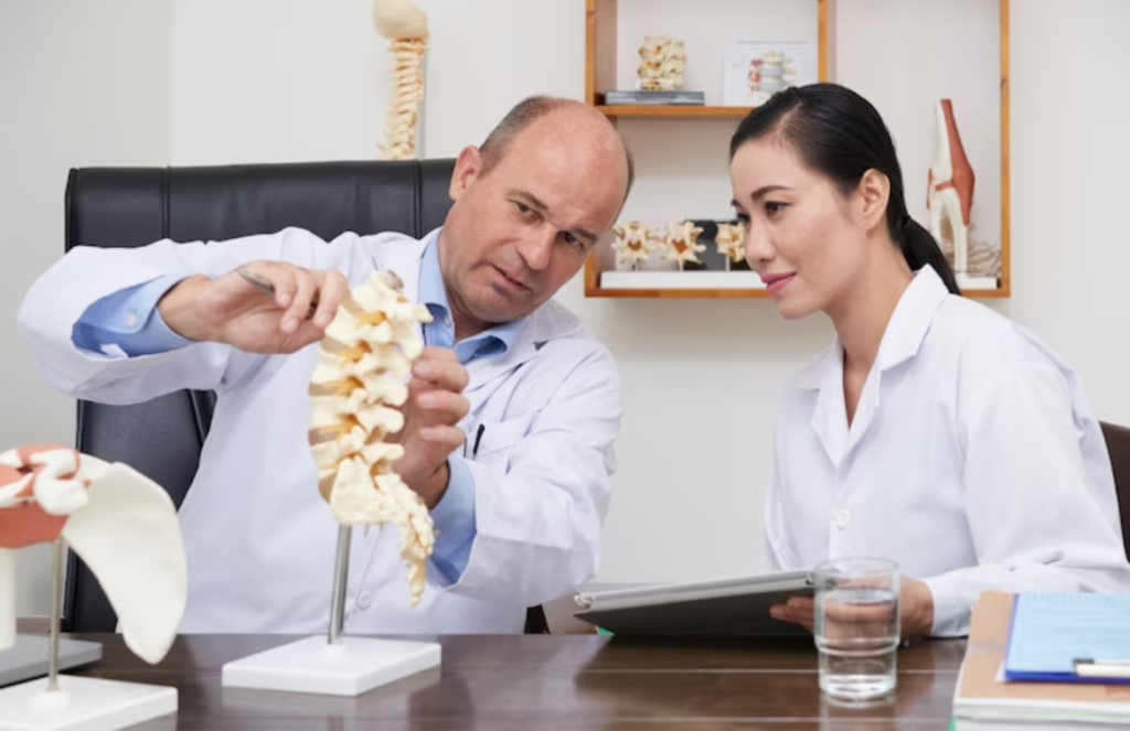
A thorough understanding of what clinicians want to know from imaging of soft tissue, bone, and bone marrow tumors does allow the radiologist to better analyze and communicate the relevant findings, making use of a common multidisciplinary language.
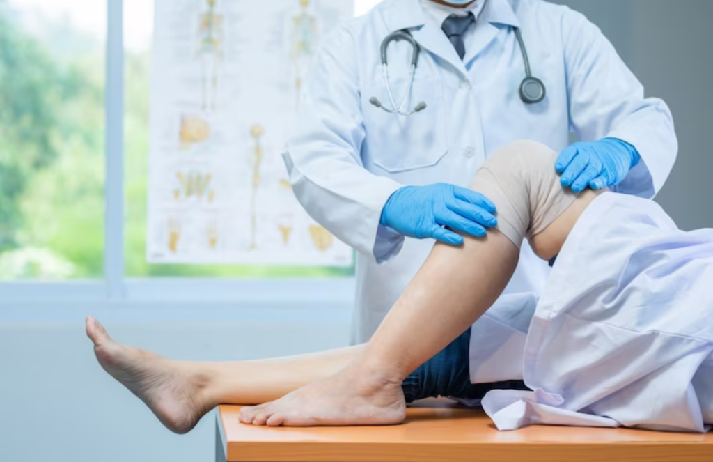
The focus is on the inclusion of a detailed patient history relative to the tumor, indicating to the clinician and the patient a clear engagement in the patient’s care and an insight into the desired imaging objectives. Clinicians prefer to know from imaging for soft tissue, bone, and bone marrow tumors.
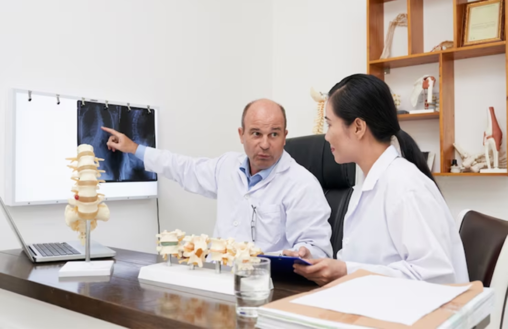
The functioning of imaging in the setting of soft tissue, bone, or bone marrow tumors relies on the capacity to accurately characterize, localize, and also quantitatively quantify the abnormality. A radiologist’s interpretation and reporting of these key imaging findings does allow for a differential diagnosis, can indeed add specificity and confidence in diagnosis, and allows for a determination regarding the need for biopsy. It also provides a shared platform from which the clinical team and patient can discuss further diagnostic management and an initial treatment plan. The appropriate characterization of a tumor can often provide insight into the potential staging of the tumor and also guide the need for additional imaging before biopsy.
Adult tumors of soft tissue, bone, and also bone marrow do encompass a broad range of histopathologies with varying immune-histochemical and molecular properties, but the tenets of imaging for such tumors and their value to clinicians and patients are consistent regardless of tumor type. This is what oncology investigations are all about.
Understanding the relevance and influence of the patient’s history and addressing the important imaging features of a tumor in a consistent and concise manner in the radiology report does elevate the perceived worth of the radiologist’s input into patient care. The focus is on magnetic resonance imaging (MRI) findings and also provides guidance on what clinicians prefer to know and how to report this information, making use of a clear and uniform approach.
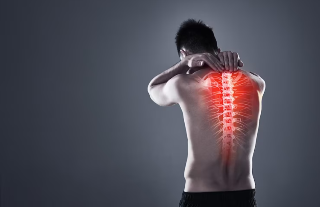
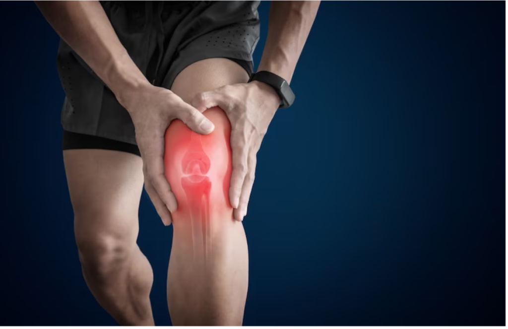
Soft tissue tumors
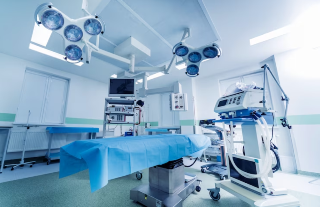
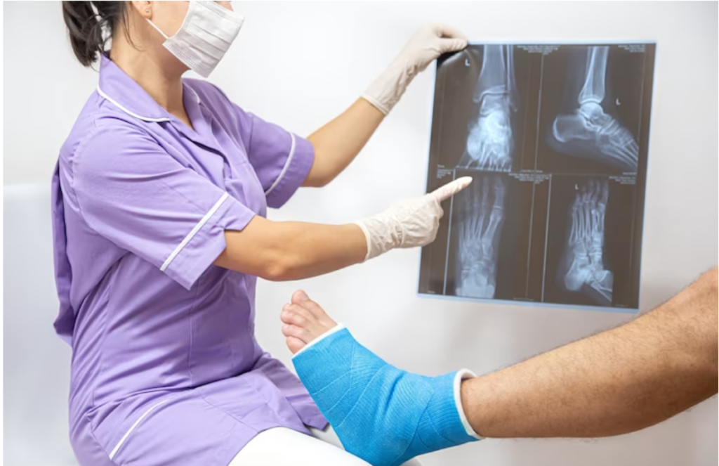
Although the majority of soft tissue tumors (STT) are indeed benign, the morbidity and mortality associated with soft tissue sarcoma (STS) are indeed important and warrant that radiologists have a fundamental knowledge of their imaging presentations, treatment methods, and goals. Radiographs, ultrasound, computed tomography (CT), and also positron emission tomography (PET)/CT can all often provide useful information for the differential diagnosis of an STT. The purpose of radiographs for the evaluation of STT is often really underestimated, but they can provide highly valuable information, like the identification and characterization of associated mineralization and also osseous lesion origin and the identification of underlying cortical involvement, which can rather influence the differential diagnosis.
MRI is the imaging technique of choice, as it often allows for a limited differential diagnosis. For instance, the most common myxoid lesions—soft tissue myxoma, myxoid liposarcoma, and myxofibrosarcoma—can usually be distinguished by their imaging characteristics in the vast majority of cases on MRI. An understanding of the relevance of the MRI features does favor a benign versus malignant diagnosis, which is paramount to making a determination of radiology-pathology concordance or discordance and also making appropriate recommendations for the next steps in management. For STS in particular, the key MRI features that tend to help predict its behavior and also potentially impact surgical management are the tumor’s anatomical site (intramuscular, subcutaneous, intra-articular, extensive/multifocal), size, depth, margin, and morphological/enhancement complexity. Specific locations can also improve diagnostic accuracy, like deep to the scapular tip for elastofibroma and hand or foot for fibromatosis. Reporting of an associated inflammatory edema-like signal or hemorrhage in the surrounding soft tissues or even associated fascial enhancement extending from the tumor (fascial tails) is also crucial, as these areas can contain tumor cells and also need to be addressed in the treatment plan to maximize local control.
Conclusion
Oncology and orthopedic issues are well investigated for better treatment.